Building Value to Science and Customers

We recently released a new application note based on the article by Anette Gjörloff Wingren’s group at Malmö University which was published in September 2014. The publication of the article was communicated in the September 2014 press release “Researchers propose PHI’s technology to assess reduction in cancer cell growth”.
Application notes are a fundamental part of marketing scientific instrumentation. By briefly describing significant applications of scientific instrumentation, application notes highlight the value to customers and ultimately the value of a of novel technology like PHI's HoloMonitor.
Flow cytometry
The article adds to the accumulating scientific evidence showing that PHI's HoloMonitor technology has the potential to supersede flow cytometry as the dominating technology for quantitative cell analysis. In 2012 the flow cytometry market was valued at 3.1 billion USD according to a market report from BCC Research.

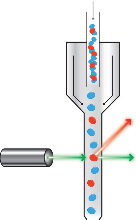
Flow cytometry (cyto = cell, metry = measure) use laser light to analyze hydraulically aligned cells.
The current form of flow cytometry was developed in the 1960s, as it proved impossible to automate microscopes without the aid of computers. Today, however, automated microscopes, powered by high speed computers, challenge the cell analysis hegemony of flow cytometers.
Flow cytometry’s principal drawbacks are that it is non-visual and not suitable to study the same cells over time. It requires that the cells are extracted from their growth environment and individually forced through a narrow tube at high speed, making flow cytometry a destructive and highly invasive analytical method.
Conventional time-lapse microscopy
To quantify cell behavior over extended periods, cell biologists are therefore compelled to use conventional time-lapse microscopy in parallel with flow cytometry. However, conventional time-lapse microscopy has several major deficiencies:
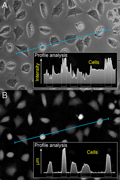
- There is no well-defined border between the cell and the background, making it difficult to automatically track and quantify cells over time non-invasively.
- To track cells accurately invasive stains are needed.
- Only a small number of cells are visible in a single field of view.
Due to these deficiencies, flow cytometry has remained the dominant cell analysis method, in spite of its flaws. As a result, cell systems are seldom observed and quantified over time. This has led to that the growth dynamics of cancer cells is poorly studied and is currently not very well understood, for example.

A profile analysis showing cell borders when imaged with conventional time-lapse microscopy (A) and HoloMonitor technology (B).
Time-lapse cytometry
Conventional time-lapse microscopy directly records an image of the cells. The PHI HoloMonitor creates images in an entirely different way. Instead the information carried by the light is recorded and the image is numerically created by a computer.
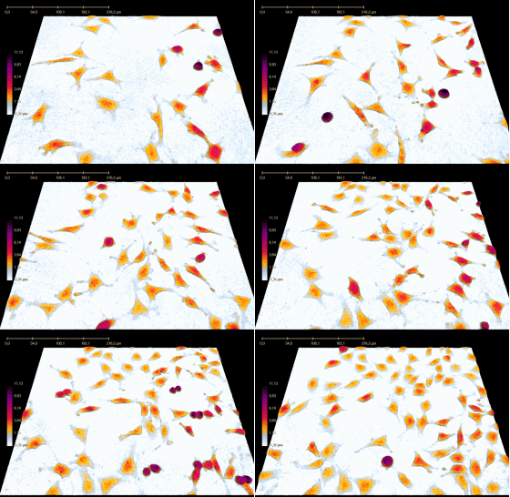
This allows HoloMonitor to visualize, track and quantify physical properties of cells over time, without the use of invasive stains. It also allows images to be digitally refocused and assembled into a time-lapse movie containing thousands of cells in each image frame. As suggested by the article authors, this enables HoloMonitor to circumvent the deficiencies of conventional time-lapse microscopy and combine the analytical benefits of time-lapse microscopy with flow cytometry in a single revolutionary device for quantitative cell analysis – a time-lapse cytometer.

Time-lapse of dividing cells, created by HoloMonitor.
References
- Press release: “Researchers propose PHI’s technology to assess reduction in cancer cell growth”
- Article: “Digital holographic microscopy for non-invasive monitoring of cell cycle arrest in L929 cells” by Falck Miniotis et al. (2014)
- Application note featuring Dr. Maria Falck Miniotis, Malmö University
- BCC Research's flow cytometry market report


