Label-Free Cell Migration Assays
The HoloMonitor® Cell Migration Assays deploy label-free live cell imaging to in vitro quantify cell migration, invasion and motility using standard culture conditions — saving precious cells and lab hours as an alternative to wound healing and transwell assays.
Cell Proliferation and Migration
Aside from rampant cell proliferation, metastatic cancer is characterized by cell migration. In laboratory experiments, cell proliferation and migration are traditionally studied separately, using distinctly different assays and protocols.
Ideally, these hallmarks of cancer should be studied simultaneously under the exact same conditions. However, this has been difficult to achieve in the past, as the established methods to assess cell migration, wound healing assay (a.k.a. scratch assay) and transwell assay (a.k.a. Boyden chamber), require cell starvation and special sample preparation.
Cell Motility — An Alternative to the Transwell Assay
Zhang et al. (below) have shown that the average cell motility provides similar cell migration results as obtained by wound healing and transwell assays. Cell motility is quantified by measuring the non-directional movement of cells in a standard cell culture sample. This makes it possible to quantify in vitro cell migration and cell proliferation simultaneously, without requiring cell starvation or special sample preparation, nor does it require any labels.

In Evaluation of Holographic Imaging Cytometer HoloMonitor M4 Motility Applications, Cytometry Part A (2018), Zhang et al. show that cell motility provides similar results as determining migration by wound healing assay.
These findings were applied in Bi-allelic Loss of CDKN2A Initiates Melanoma Invasion via BRN2 Activation, Cell Cancer (2018).
Yuntian Zhang, Ph D Postdoctoral Scholar
University of California, San Francisco
Quantifying Cell Motility
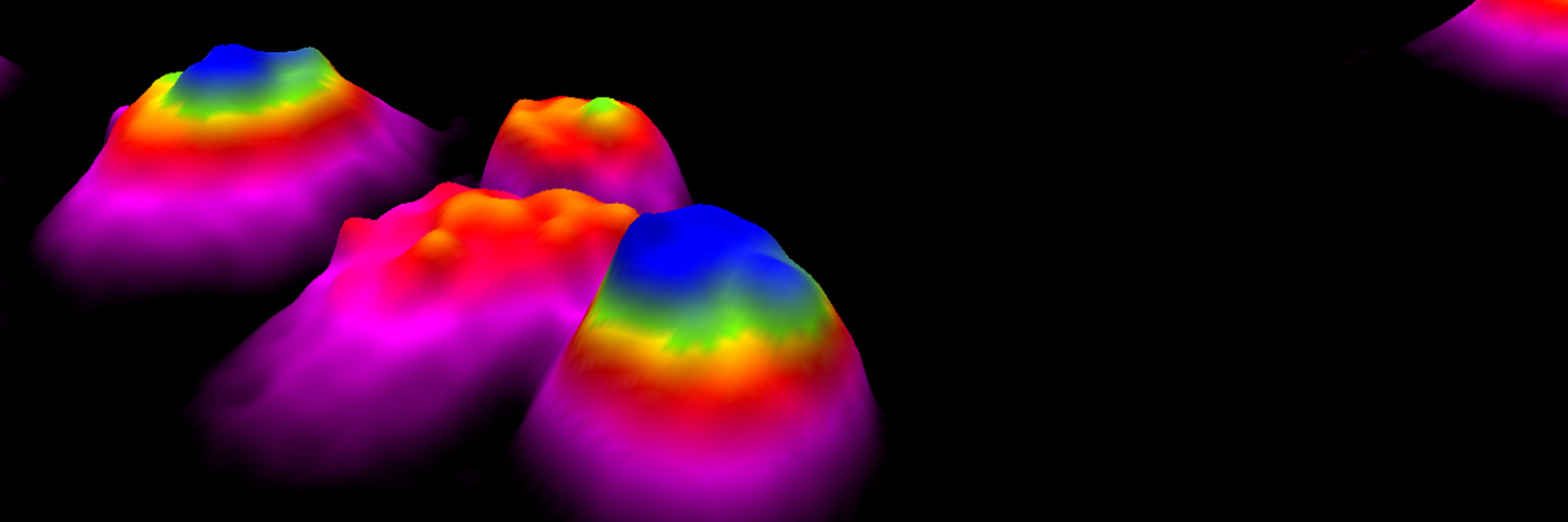
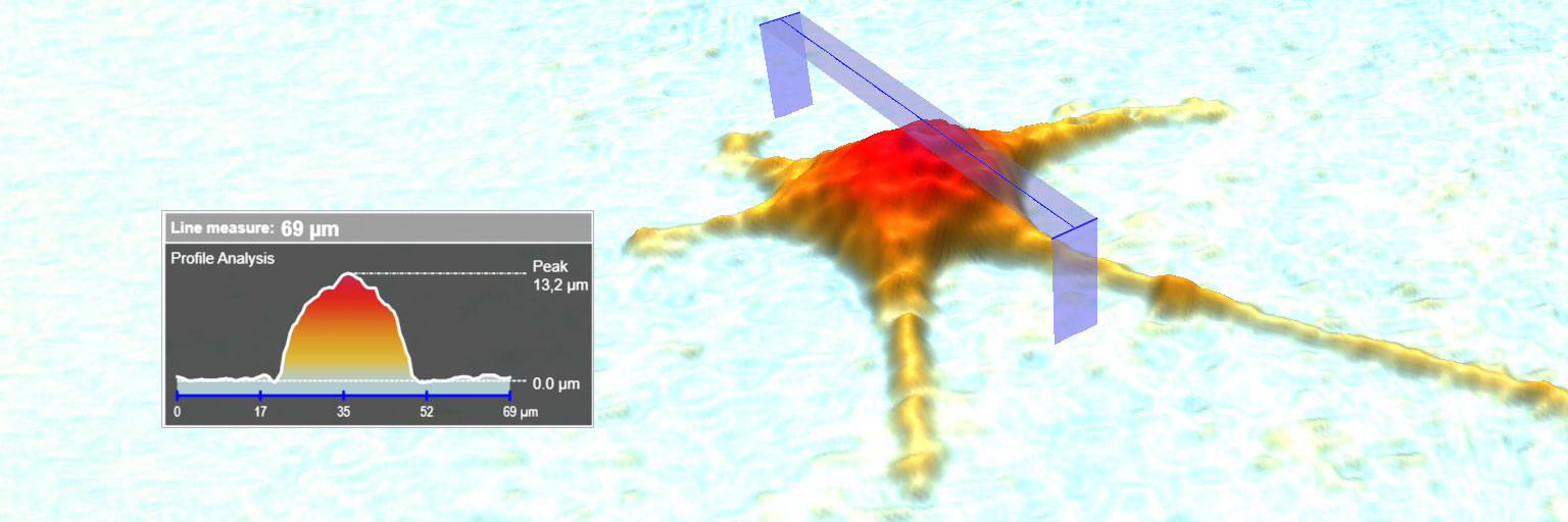
Using HoloMonitor®, label-free cell motility data is gently obtained by recording a time-lapse image sequence of proliferating cells. The mean cell motility is automatically determined by applying the HoloMonitor Cell Motility Assay on the recorded time-lapse sequence, while individual cell motility data is obtained by tracking selected cells using HoloMonitor Cell Tracking.
As their is no need to specifically prepare the cells to quantify motility, cell proliferation data can be obtained as well by applying the HoloMonitor Cell Proliferation Assay on the same time-lapse image sequence, without requiring any additional samples or lab work.
A Novel Cell-Friendly Approach Saving Precious Cells
Conceptually, the HoloMonitor Assays operate differently than conventional assays. When using conventional assays, the cells are prepared according to and specifically for the selected assay, resulting in that the cells can seldom be used for any other assay.
When working with HoloMonitor, the cells are generically prepared and imaged. After recording, one or more HoloMonitor assays are applied on the time-lapse images to obtain multiple results from the same cell sample — for example cell migration and cell proliferation data.
Indeed, this technique has been demonstrated to accurately measure cell growth, cell motility and migration, cell death, cell cycle, and drug responses in a wide variety of cell lines in the laboratory and in the clinical setting.
Laura V. Croft et al.
This novel approach reduces the necessary amount of lab work. But, more importantly, it facilitates the use of precious primary cells over cell lines to improve clinical relevance by dramatically reducing the number of primary cells needed to obtain results.
Classic Wound Healing Assay
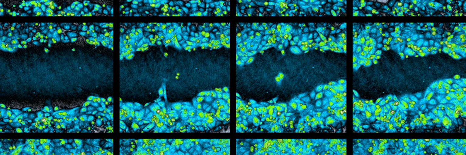
When there are cells to available and when a firmly established assay is preferred, the label-free HoloMonitor Assays also include the HoloMonitor Wound Healing Assay. In addition to the assay, cell tracking may be applied on selected cell front cells for detailed individual cell movement and morphology analysis.

The HoloMonitor live cell imaging system performing a cell migration assay.


