
The HoloMonitor Blog
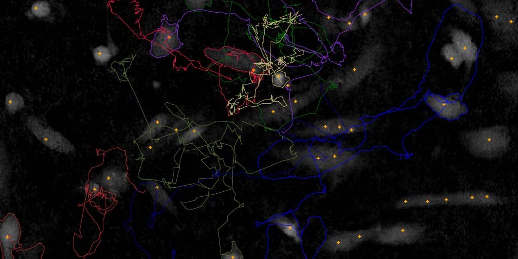
A study of cancer cells by digital holographic imaging, fluorescence and a combination thereof
Digital holographic imaging is a label-free, non-toxic quantitative method that can be used for single cell analysis for a long time period. Sofia Kamlund, PhD, Lund University and Phase Holographic Imaging, has used this method in her thesis Not all those who wander are lost – A Study of Cancer Cells by Digital Holographic Imaging, Fluorescence and a combination there of. The technique allows the researcher to follow changes over time and evaluate cell response on living cells.
Learn more …

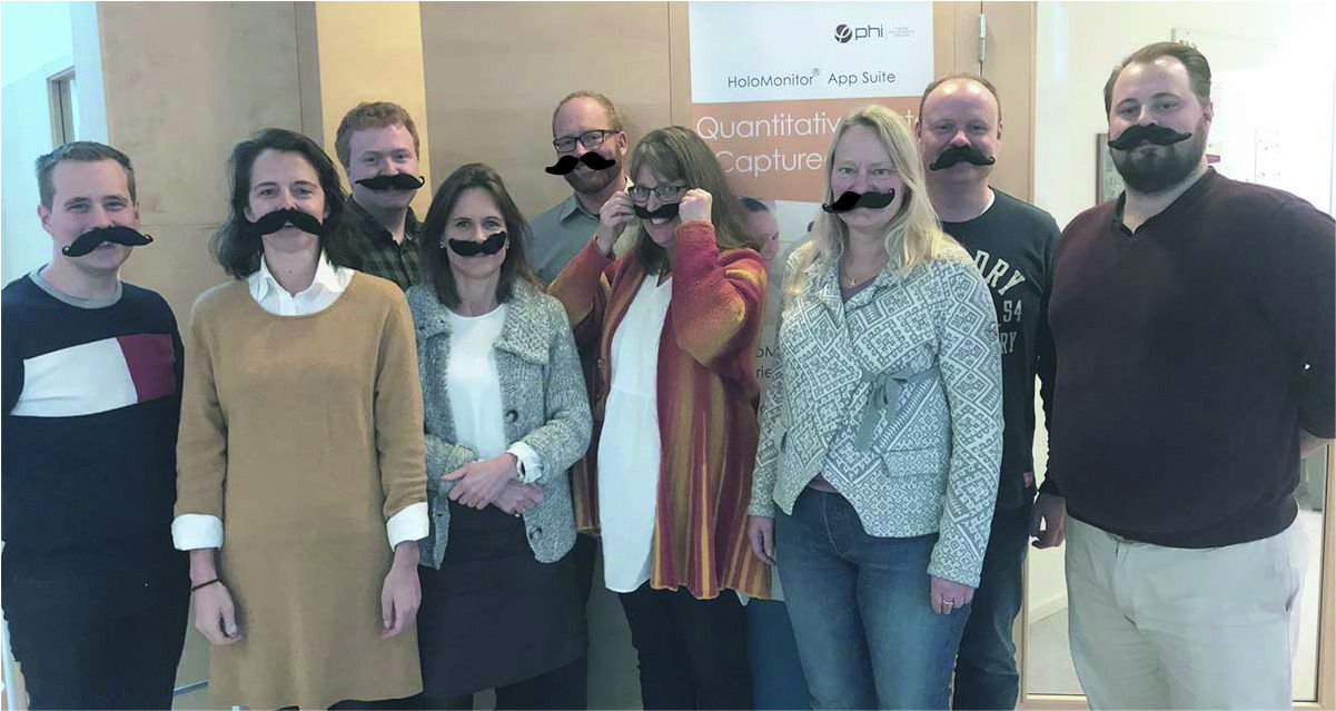
A Change of Measure – HoloMonitor Distributor Meeting
Learn more …
Prostate Cancer Research in Movember
Globally, prostate cancer is the second most commonly occurring cancer in men and the fourth most commonly occurring cancer overall. This video shows Etoposide treated DU 145 prostate cancer cells undergoing programed cell death, apoptosis.
Learn more …
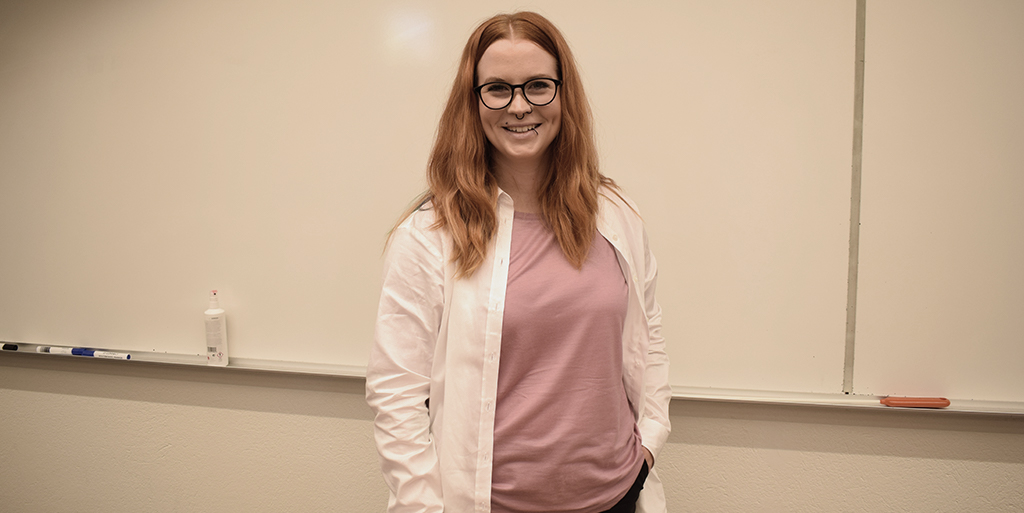
Dissertation: A study of Cancer Cells by Digital Holographic Imaging
Sofia Kamlund has studied Cell Division and Cell Movement using Digital Holographic Microscopy. Her research presents new ways of analyzing individual cells by using Holographic Microscopy alone or in combination with Fluorescence Microscopy.
Learn more …
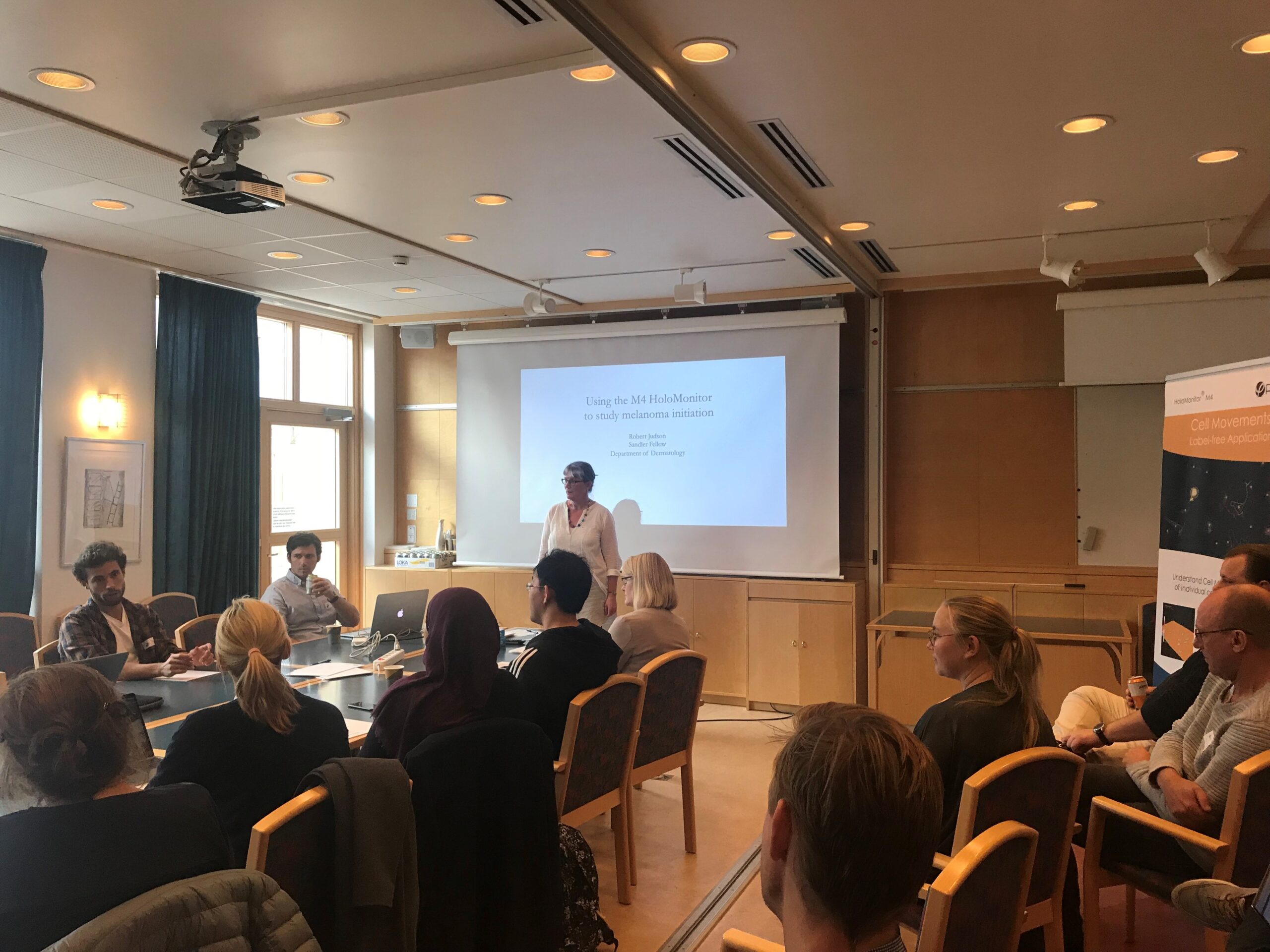
HoloMonitor User Meeting
PHI had a successful Holomonitor User Meeting where we discussed how to use HoloMonitor with focus on cancer research. The presentations showed new methods within cancer research and cell biology.
Learn more …
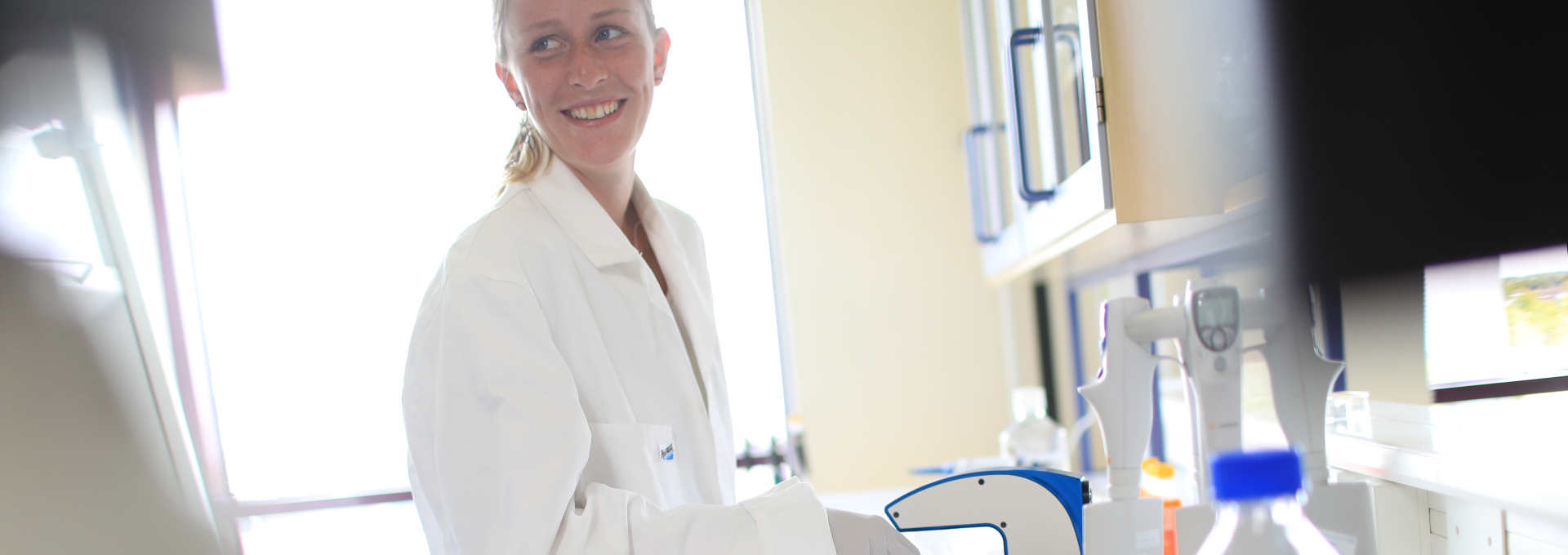
Best Poster Prize to Louise Sternbaeck
PHI’s Industrial Doctoral Student Louise Stenbaeck received a prize from Danish Cancer Society for her poster “Holographic microscopy: Macrophage-uptake of SA-MIPs”.
Learn more …